
save
$25.01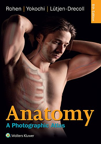
Anatomy: A Photographic Atlas, 8th Edition
Prepare for the lab and OR with Anatomy: A Photographic Atlas, 8th Ed — over 1,200 cadaver photographs in color. Get your digital copy today.
Want to preview before you buy? Click Request Free Sample and tell us which chapter or pages you’d like to see, along with your email address. We’ll send you those pages from the eBook so you can be sure it’s the right one before buying.
- Instant download & lifetime access Available as soon as your payment is confirmed. Your download link is also sent to the email address you provide at checkout, and you can download your purchased eBook anytime from My Account → Downloads with unlimited downloads. An account is created automatically using the same email address.
- Own your eBook Receive the available PDF/EPUB file directly after purchase. No rental or temporary access — download and keep your eBook for personal use with lifetime access.
- Full refund guarantee If the eBook you receive does not match the details and description provided on the product page, contact [email protected] and we’ll issue a full refund within 24 hours.
- Secure payment through PayPal Pay securely by debit or credit card or with your PayPal account. Your card details are processed by PayPal and are never stored on our site.
$19.99$45.00
The Problem This Book Solves
Textbook illustrations and diagrams can only go so far when you are standing in an anatomy lab or preparing for a surgical procedure. The gap between idealized drawings and the messy, three-dimensional reality of the human body is vast. Anatomy: A Photographic Atlas bridges that gap by placing real cadaver dissections at the center of your study.
This eighth edition delivers over 1,200 full-color photographs, each one meticulously labeled and organized by body region. Whether you are a medical student dissecting for the first time or a resident refining your surgical knowledge, this atlas eliminates the abstraction and shows you what actual human anatomy looks like.
About Anatomy: A Photographic Atlas, 8th Edition
Anatomy: A Photographic Atlas (Eighth Edition) is a comprehensive visual guide to human anatomy created by Johannes W. Rohen, Chihiro Yokochi, and Elke Lütjen-Drecoll. Published by Schattauer GmbH and Wolters Kluwer, this edition features 1,209 figures — 1,096 in color — plus 113 radiographs, CT scans, and MRI images that correlate dissection views with clinical imaging.
Unlike traditional illustrated atlases, every photograph in this book comes from actual cadaver specimens. The dissections are performed with exceptional care to preserve natural relationships among organs, muscles, nerves, and vessels. The result is a reference that looks and feels like the real thing — exactly what you need to recognize structures in the lab and in the operating room.
Who Is This Book For?
This atlas is designed for anyone who needs a reliable, photograph-based reference for human anatomy. It is especially valuable for:
- Medical students enrolled in gross anatomy courses who must dissect and identify structures under time constraints
- Surgical residents and fellows who require a fast, accurate visual refresher before entering the OR
- Anatomy instructors and professors who want to show students realistic dissections without the odors and logistics of a wet lab
- Physician assistants, nurse practitioners, and physical therapists who need to apply anatomical knowledge in clinical practice
- Radiology technicians and medical imaging professionals who will benefit from the side-by-side CT/MRI correlations
Key Takeaways
Working through this atlas, you will master the following essential areas of human anatomy:
- Regional musculoskeletal anatomy — every muscle, bone, joint, and ligament photographed in natural position
- Thoracic and abdominal organ topography — precise relationships of the heart, lungs, liver, intestines, and kidneys
- Neuroanatomy of the brain and spinal cord — detailed cross-sections and dissections of cranial nerves and white-matter tracts
- Vascular anatomy of the head, neck, and extremities — arteries, veins, and lymphatic drainage shown with clarity
- Radiographic anatomy — 113 radiographs, CT scans, and MRI images that bridge dissection views to clinical imaging
- Pelvic and perineal anatomy — male and female reproductive organs, pelvic floor, and urogenital triangles
- Surface anatomy and palpation landmarks — photographs that help you translate internal structures to external landmarks
- Systematic dissection sequences — layered dissections that show how to approach cadaveric exploration step by step
Why Choose This Edition
Most anatomy atlases rely on hand-drawn illustrations or computer-generated renderings. While these can be beautiful, they often simplify or idealize structures. Anatomy: A Photographic Atlas uses real photographs — so what you study is exactly what you will encounter in the lab.
Compare this to Netter’s Atlas of Human Anatomy, which uses classic illustrations, or Grant’s Atlas of Anatomy, which combines drawings with some photographs. The Rohen atlas is unique in its commitment to photography. Every region is presented with multiple views, including deep dissections that reveal layers other books skip. The inclusion of over 100 radiographs, CTs, and MRIs also gives you a direct link from dissection to diagnostic imaging — a feature that is especially valuable for board exams and clinical rotations.
Furthermore, the eighth edition has been updated with improved labeling and expanded imaging correlations. It remains the gold standard for photograph-based anatomy education.
About the Author
Johannes W. Rohen is a distinguished anatomist whose career has been dedicated to the study of human morphology. He has authored multiple editions of this atlas and is recognized worldwide for his contributions to anatomical education. Chihiro Yokochi and Elke Lütjen-Drecoll bring decades of experience in anatomy and ophthalmology, respectively, ensuring that the dissections are both accurate and pedagogically sound.
The publisher, Schattauer GmbH in partnership with Wolters Kluwer, is a leading name in medical and scientific publishing. Wolters Kluwer’s imprint (Lippincott Williams & Wilkins) is trusted by medical schools and hospitals globally. This combination of expert authorship and institutional backing gives the atlas a level of authority that cannot be matched by self-published or lesser-known resources.
Final Assessment
Absolutely. If you are serious about learning human anatomy from a real-world perspective, this atlas is an indispensable tool. The photographs are not just beautiful — they are pedagogically effective. Students who use this atlas consistently report higher confidence in the dissection lab and better retention of anatomical relationships.
For the price of a single textbook, you gain access to a lifetime-reference that will serve you from your first anatomy exam through your surgical boards. The eighth edition is current, well-organized, and packed with learning aids that no other photographic atlas offers.
Start Reading Anatomy: A Photographic Atlas, 8th Edition Today
Whether you are a first-year medical student facing your first dissection, a surgical resident preparing for a case, or an instructor looking for the best visual aid for your students, Anatomy: A Photographic Atlas is the right choice. This eighth edition from Schattauer/Wolters Kluwer gives you the most realistic, photograph-based anatomy reference available.
Don’t settle for illustrations that don’t match reality. Add this atlas to your cart now and start studying anatomy the way it truly looks — on the cadaver and in the clinic.
Related products

Essentials of Epidemiology in Public Health, Fourth Edition
$25.99Master epidemiologic methods with this essential Fourth Edition textbook from Jones & Bartlett Learning, perfect for public health students and professionals.
$78.00-
Anthony’s Textbook of Anatomy & Physiology 18th Edition – Mosby
$20.00Master human anatomy and physiology with this clear, comprehensive 18th Edition from Mosby. Perfect for nursing and allied health students.
$62.00 -
Epilepsy: A Queen Square Textbook (First Edition) – Comprehensive Adult Epilepsy Guide
$40.99The authoritative first edition from Queen Square experts—your essential resource for mastering epilepsy diagnosis and management.
$105.00 -
Antibiotic Drug Resistance (1st ed.)
$64.99Comprehensive guide to antibiotic drug resistance mechanisms, clinical impact, and solutions. Ideal for researchers and clinicians. Order now.
$185.00 -
Child and Adolescent Mental Health Screening, Evidence-Based Assessment, Intervention, and Health Promotion, 3rd Edition
$30.99The essential evidence-based guide for child and adolescent mental health screening, assessment, and intervention—updated third edition with practical tools and KySS framework.
$65.00 -
Vascular Surgical Emergencies: Well-Prepared for Duty
$28.00Master 50 vascular surgical emergencies with proven diagnostic and therapeutic protocols. The 2026 Springer guide from Susanne Regus — essential for every vascular surgeon.
$49.00 -
Essentials of Neuro-Psychiatry: A Classic Textbook of Nervous and Mental Disorders (1945)
$29.99Own a piece of medical history with this 1945 neuropsychiatry classic from Lea & Febiger. Essential for collectors and scholars. Add to cart today.
$60.00 -
Vascular Access: Principles and Practice 5th Edition – Comprehensive Textbook
$48.99Master all aspects of vascular access for hemodialysis, chemotherapy, nutrition, and critical care with this practical 5th edition textbook by Samuel Eric Wilson. Get the definitive how-to guide now.
$141.00