
save
$3.01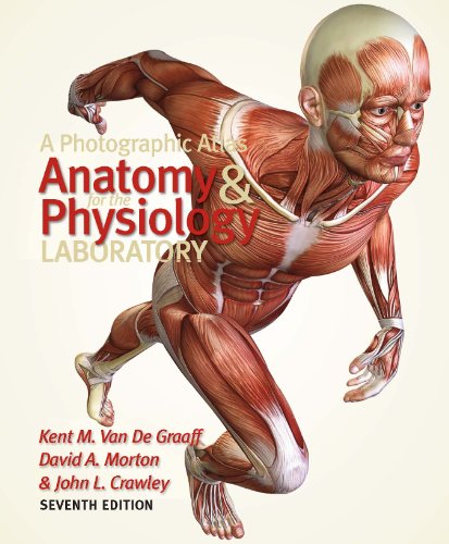
A Photographic Atlas for the Anatomy and Physiology Laboratory, 7th Edition
Get the essential photographic atlas for your anatomy and physiology lab with clear, full-color images of every body system. Perfect for lab study and dissection. Order now!
$7.99$11.00
The Problem This Book Solves
Learning human anatomy from diagrams alone is like navigating a city with a map that shows only streets, not the buildings. Medical and nursing students need to see real tissue, real organs, and real dissection to truly understand the body’s architecture. This atlas bridges the gap between textbook line art and the cadaver lab, providing high-resolution photographs that mirror what you will encounter in actual dissection.
A Photographic Atlas for the Anatomy and Physiology Laboratory, 7th Edition at a Glance
This seventh edition of A Photographic Atlas for the Anatomy and Physiology Laboratory is a visual reference designed to accompany any human anatomy or integrated anatomy and physiology course. Published by Morton Publishing Company in 2011, the atlas contains hundreds of full-color photographs of muscle, skeletal, and organ systems. Each image is carefully labeled to help you identify structures quickly and accurately. The atlas is spiral-bound for easy use on the lab bench, making it a durable companion for dissection sessions.
Who Is This Book For?
This atlas is ideal for:
- Undergraduate anatomy and physiology students
- Nursing and allied health program students
- Pre-medical and pre-dental students
- Physical therapy and exercise science majors
- Anyone taking a human anatomy lab course
- Instructors seeking a reliable visual teaching aid
Skills You Will Gain
By using this photographic atlas, you will master:
- The skeletal system — bones, markings, and joints from every angle
- The muscular system — origin, insertion, and action of major muscles
- The cardiovascular system — heart, vessels, and circulation pathways
- The respiratory system — lungs, trachea, and associated structures
- The digestive system — organs from mouth to anus
- The nervous system — brain, spinal cord, and peripheral nerves
- The reproductive and urinary systems — detailed photographic views
Why This Book Stands Out
Unlike many lab manuals that rely on simplified illustrations, this atlas uses real photographs from actual cadaver dissections. The images show the natural texture, color, and spatial relationships that drawings cannot replicate. Each photograph is accompanied by clear leader lines and labels that make identification intuitive. The atlas covers all major body systems, and its spiral binding allows it to lay flat open on the lab bench. Competing atlases often use older, lower-resolution images; this edition updated numerous photographs for greater clarity and accuracy.
Who Wrote This Book
The authors — Kent M. Van De Graaff, David A. Morton, and John L. Crawley — are experienced anatomy educators who have taught thousands of students. Their combined expertise ensures that every image and label reflects current anatomical terminology. Morton Publishing Company specializes in anatomy and physiology educational resources, further confirming the atlas’s reliability for academic use.
Final Assessment
Yes. For any student enrolled in a human anatomy lab, this atlas is an indispensable tool. The photographic quality saves hours of confusion during dissection and study. It reinforces lecture concepts with real-world visuals, and it is compact enough to carry to every lab session. While the seventh edition is not the newest, its content remains highly relevant for foundational anatomy courses. The price is a fraction of what you would pay for a full textbook, making it a cost-effective investment in your academic success.
Start Reading A Photographic Atlas for the Anatomy and Physiology Laboratory, 7th Edition Today
Stop struggling with abstract diagrams. Order your copy of A Photographic Atlas for the Anatomy and Physiology Laboratory, 7th Edition, now and gain the visual confidence you need to excel in your lab practicals. This Morton Publishing atlas is the trusted companion for students across the country. Add it to your cart and start learning anatomy the way it was meant to be seen — in vivid, real photography.
Related products

Vascular Surgical Emergencies: Well-Prepared for Duty
$22.99Master 50 vascular surgical emergencies with proven diagnostic and therapeutic protocols. The 2026 Springer guide from Susanne Regus — essential for every vascular surgeon.
$40.00-
HESI Admission Assessment Exam Review 4th Edition: Complete Study Guide for Nursing Entrance Exams
$18.99Master the HESI Admission Assessment Exam with Elsevier’s official 4th Edition study guide. Covers math, science, reading, grammar & vocabulary with practice tests.
$38.00 -
Endurance Sports Nutrition 3rd Edition – Fueling for Peak Performance
$24.99Fuel your best performance with Endurance Sports Nutrition. This science-backed guide helps you train longer, recover faster, and avoid injury with personalized fueling strategies.
$54.00 -
Glandular Physiology and Therapy: AMA Symposium Ebook
$29.99Gain rare insight into early endocrinology with this authoritative AMA symposium. Essential for medical historians and hormone therapy researchers. Buy the ebook today.
$60.00 -
Guide to the Most Common Internal Medicine Workups and Diseases: An Evidence-Based Hospital Reference
$33.99Master the most common hospital-based internal medicine workups with this evidence-based guide. Get the structured clinical reference you need for daily practice.
$65.00 -
Botulinum Toxin Procedures: A Practical Guide to – eBook
$12.99Master Botulinum Toxin Procedures with this practical guide. Step-by-step instructions, anatomy, and patient selection for safe, effective treatments.
$21.00 -
Davis’s Comprehensive Handbook of Laboratory & Diagnostic Tests, 8th Edition
$26.99Master every lab and diagnostic test with nursing implications you can trust. The 8th edition of Davis’s Comprehensive Handbook is your pocket-sized guide to accurate interpretation and safe patient care.
$56.00 -
Essentials of Neuro-Psychiatry: A Classic Textbook of Nervous and Mental Disorders (1945)
$29.99Own a piece of medical history with this 1945 neuropsychiatry classic from Lea & Febiger. Essential for collectors and scholars. Add to cart today.
$60.00