save
$25.00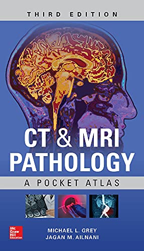
CT & MRI Pathology: A Pocket Atlas, 3rd Edition
Instant access to the essential pocket atlas for CT and MRI pathology interpretation — perfect for radiology residents and clinicians seeking rapid, accurate diagnosis.
Want to preview before you buy? Click Request Free Sample and tell us which chapter or pages you’d like to see, along with your email address. We’ll send you those pages from the eBook so you can be sure it’s the right one before buying.
- Instant download & lifetime access Available as soon as your payment is confirmed. Your download link is also sent to the email address you provide at checkout, and you can download your purchased eBook anytime from My Account → Downloads with unlimited downloads. An account is created automatically using the same email address.
- Own your eBook Receive the available PDF/EPUB file directly after purchase. No rental or temporary access — download and keep your eBook for personal use with lifetime access.
- Full refund guarantee If the eBook you receive does not match the details and description provided on the product page, contact [email protected] and we’ll issue a full refund within 24 hours.
- Secure payment through PayPal Pay securely by debit or credit card or with your PayPal account. Your card details are processed by PayPal and are never stored on our site.
$24.00$49.00
The Gap This Book Fills
Interpreting cross-sectional imaging requires a rapid, accurate connection between radiographic findings and underlying pathology. Without a concise visual reference, radiologists, residents, and clinicians spend precious time flipping through dense textbooks or searching fragmented online resources. CT & MRI Pathology: A Pocket Atlas eliminates this inefficiency by delivering a focused, image-rich guide that matches common pathologies to their CT and MRI appearances — exactly what you need at the reading station or on rounds.
What Is CT & MRI Pathology: A Pocket Atlas?
CT & MRI Pathology: A Pocket Atlas, 3rd Edition (McGraw-Hill Professional, 2018) is a compact, image-driven reference designed for rapid identification of pathology on computed tomography and magnetic resonance imaging. Co-authored by Michael L. Grey and Jagan Mohan Ailinani, this pocket-sized atlas organizes over 400 high-quality images by body system, pairing each radiographic finding with a concise description of the underlying disease process. The eText edition provides instant digital access on any device, making it an indispensable tool for busy clinical environments.
Is This Book Right for You?
This atlas is tailored for radiology residents preparing for board exams, practicing radiologists needing a quick refresher, and clinicians (neurologists, orthopedists, oncologists) who routinely interpret cross-sectional images. It also benefits medical students rotating through radiology and technologists seeking to understand the pathology behind the scans they acquire. If your work involves CT or MRI interpretation and you value portability without sacrificing depth, this book belongs in your digital library.
Skills You Will Gain
- Systematic pathology recognition — from brain tumors to spinal trauma, each region is covered with correlative imaging
- Distinguishing features for common versus rare pathologies, helping you narrow differentials faster
- Normal variants and pitfalls that mimic disease, reducing false-positive interpretations
- Key imaging findings for inflammatory, neoplastic, vascular, and degenerative conditions
- Pattern-based approach to interpreting CT and MRI signal characteristics (e.g., T1, T2, contrast enhancement)
- Practical annotation of each image with arrows and labels for quick visual reference
- Updated content reflecting current diagnostic criteria and imaging protocols as of the 3rd edition
Why Choose This Edition
Unlike larger reference tomes such as Radiology Review Manual or Primer of Diagnostic Imaging, this atlas focuses exclusively on pathology rather than anatomy or technique. It is intentionally sized for the pocket — digital or print — so you can consult it during a case rather than at a desk. Competitor pocket guides often lack the image quality or disease breadth found here; the 3rd edition adds updated pathology coverage and improved image resolution. For residents who need to correlate histology with imaging quickly, this atlas bridges the gap between textbook theory and real-world interpretation better than any comparable product.
Author & Publisher Credentials
Michael L. Grey is a recognized expert in neuroradiology and head-and-neck imaging, with decades of teaching experience at leading academic institutions. Co-author Jagan Mohan Ailinani brings subspecialty expertise in musculoskeletal and body imaging. Together, they have curated a collection of cases that reflects the most clinically relevant pathology encountered in daily practice. Published by McGraw-Hill Professional — a trusted name in medical education for over a century — this atlas undergoes rigorous peer review and editorial oversight, ensuring accuracy and reliability. The 3rd edition (2018) incorporates feedback from residents and practicing radiologists, making it a current, authoritative resource.
Is It Worth It?
For radiology trainees and busy clinicians, this atlas delivers exceptional value. Its concise format, high-quality images, and pathology-focused organization directly address the daily challenge of rapid image interpretation. The digital edition offers portability across devices, searchability, and always-available access — advantages that a printed book cannot match. Priced competitively against other pocket atlases and digital textbooks, the 3rd edition is a cost-effective investment that will pay dividends in improved diagnostic confidence and faster study sessions. We consider it an essential addition to any radiologist’s digital toolkit.
Add CT & MRI Pathology: A Pocket Atlas, 3rd Edition – eBook to Your Library
Stop wasting time hunting for pathology correlations. With the 3rd edition eText from McGraw-Hill Professional, you gain immediate, lifetime access to a curated visual library of CT and MRI pathology. Whether you are a resident cramming for boards or a clinician interpreting scans after hours, this pocket atlas puts the answers at your fingertips. Order your digital copy today and start interpreting with confidence — no waiting, no shipping, just instant knowledge.
Related products

EMDR Therapy Third Edition: Basic Principles, Protocols, and Procedures by Francine Shapiro
$23.99Master EMDR therapy with this definitive third edition by Francine Shapiro. Learn the complete eight-phase protocol for treating PTSD and trauma effectively.
$44.00-
Neuroanatomy: An Illustrated Colour Text 6th Edition
$19.99Master neuroanatomy with this visual, concise Sixth Edition by Crossman & Neary. Includes enhanced eBook with bonus figures and self-assessment.
$35.00 -
Anthony’s Textbook of Anatomy & Physiology 18th Edition – Mosby
$20.00Master human anatomy and physiology with this clear, comprehensive 18th Edition from Mosby. Perfect for nursing and allied health students.
$62.00 -
Feigin & Cherry’s Textbook of Pediatric Infectious Diseases, 9th Edition
$94.99Get the definitive 9th edition of Feigin and Cherry’s Textbook of Pediatric Infectious Diseases – the premier two-volume reference for pediatric ID professionals. Order now for instant digital access!
$271.00 -
Farquharson’s Textbook of Operative General Surgery 11th Ed
$40.99Master operative surgery with the Eleventh Edition of Farquharson’s essential textbook for surgical trainees, featuring 490 color illustrations and expert guidance.
$91.00 -
Guide to the Most Common Internal Medicine Workups and Diseases: An Evidence-Based Hospital Reference
$30.99Master the most common hospital-based internal medicine workups with this evidence-based guide. Get the structured clinical reference you need for daily practice.
$59.00 -
General Thoracic Surgery, 7th Edition
$59.99Get the 7th edition General Thoracic Surgery eBook – the comprehensive two-volume guide to all open and endoscopic thoracic surgical procedures.
$149.00 -
Child and Adolescent Mental Health Screening, Evidence-Based Assessment, Intervention, and Health Promotion, 3rd Edition
$30.99The essential evidence-based guide for child and adolescent mental health screening, assessment, and intervention—updated third edition with practical tools and KySS framework.
$65.00